“Todo hombre puede ser, si se lo propone, escultor de su proprio cerebro” (Every man can be, if he so desires, the sculptor of his own brain). – Santiago Ramón y Cajal
Here’s a fun cocktail party fact for you: the winner of the 1906 Nobel Prize in Physiology or Medicine was also an artist. And an immensely talented one.
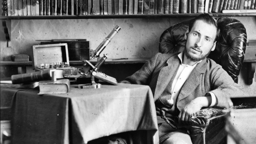
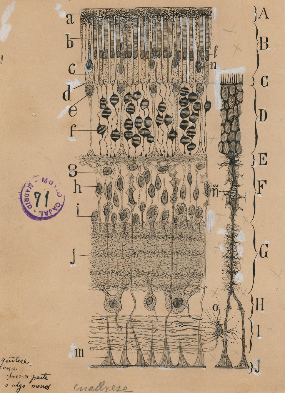
Spanish neuroanatomist Santiago Ramón y Cajal (1852-1934), considered the father of modern neuroscience, created some of the most spectacular renderings of the brain you’ll ever see. Exhibit #1, immediately below, is his depiction of the layers of the retina, one of many drawings included in a traveling exhibition of Cajal’s work. It’s the first time they’ve been presented here in the United States; the show is currently running in New York. The level of detail–the stippling and cross-hatching, the fine lines–is exquisite. If you go to Google Images and type in ‘retinal layers’, you’ll be presented with innumerable depictions, most of them very colorful, many with catchy captions, but none–for me, anyway–as beautiful as Cajal’s. All the more impressive that he produced his image in 1904, with pencil and ink. No computer, no Adobe software, no bells and whistles.
Cajal shared the 1906 Nobel with Camillo Golgi; the men were recognized for their work on elucidating the structure of the nervous system. Golgi, a histologist, developed the tissue stain–a solution of silver nitrate–that enabled Cajal’s observations. (Golgi called it la reazione nera, or “the black reaction”).
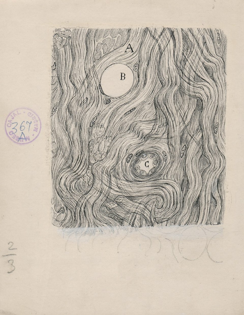
Here’s another gem:

Science journalist Joanna Klein speculated about some of Cajal’s possible artistic influences; she points out that the glial cells in the upper left hand corner of this image resemble the unhappy man in Edvard Munch’s “The Scream”.
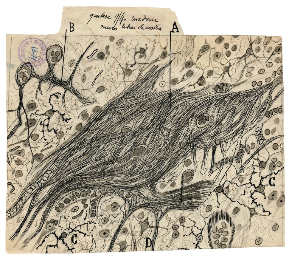

And while I, for one, sense the influence of van Gogh in this image, I may be channeling my last post.
Trivia tidbit: In honor of Cajal, the spring 1998 Columbia Space Shuttle mission had microscope slides and drawings from the Cajal Institute in its payload. The 16-day flight, called Neurolab, was dedicated to the advancement of neuroscience research, investigating effects of microgravity on the brain and nervous system. Cajal also has an asteroid (117413) named in his honor.
And one more thing: For some images that are in many ways antithetical to Cajal’s, check this out.

I very much enjoyed this post and forwarded it to my APS roommate who practices in Rome, Italt. Her blog is called, “Stethoscope on Rome”. She is a terrific story-teller!
Anne
LikeLike
Neat. I draw when designing my quilts, but the drawings aren’t very artistic! I always wanted to draw better than I do.
LikeLike
I love the most wonderful way your blogs lead one to another. Not only this one today, but Brainbow and left Brain right Brain As well. Thanks.
LikeLike
Much appreciated, Steve; glad you’re enjoying the posts!
LikeLike
These images are absolutely stunning! Amazing what he did free hand. I have seen the brainbow images before (maybe a Harvard Magazine lying around) but it’s been a while. Thank you for bringing color and beauty into our lives during the dead of winter
LikeLiked by 1 person
I think the exhibit in NYC actually includes one or two of the Brainbow images–or some similar contemporary shots–as points of comparison. Thanks, as always, for stopping by!
LikeLike
Stunning! I wonder how much these beautiful renderings of detailed structure influenced scientists’ research into function. I’ll have to ask my father (ophthalmologist), if he’s seen the drawing of the retina. As always, thank you, Jeanne, for sharing. – Vince
LikeLiked by 1 person
Vince, I wouldn’t be at all surprised if Cajal’s drawings were included in the textbooks your father used when he was in medical school. It sounds as though they were considered THE definitive images of the retina well into the 1960’s. Thanks for stopping by!
LikeLike
Extraordinary. Thanks J. Classic Augenblick.
LikeLiked by 1 person